What are the types of dog eyelid tumors?
Tumors of the eyelid may be benign (i.e. non-cancerous) or malignant (i.e. cancerous). Thankfully, about 75% of dog eyelid tumors are benign. And even those that are technically malignant may sometimes still act in a more benign fashion.
As I mentioned, tumors arise from cells in the area. This means that, broadly speaking, dogs may have masses of the meibomian glands, the haired eyelid skin, the conjunctiva, and the eyelid margins. More specifically, dogs can have the following types of eyelid tumors:
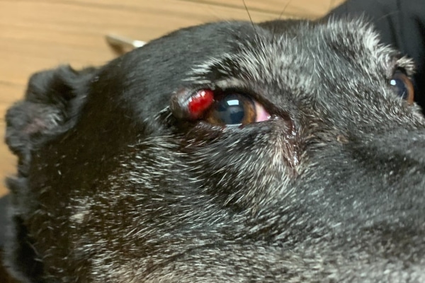
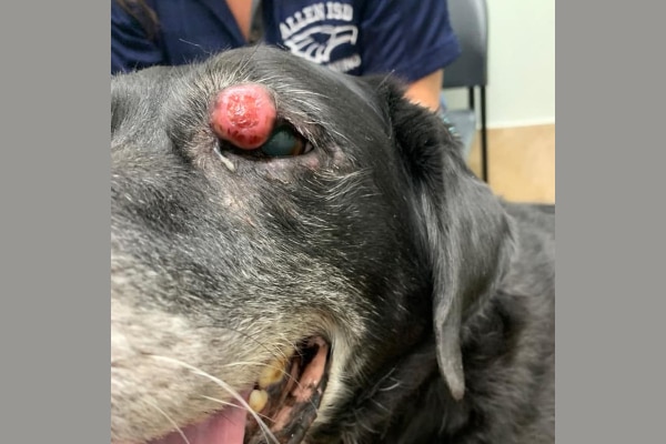
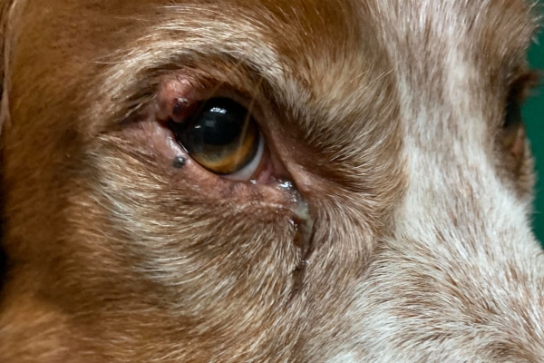
The most common eyelid tumor of older dogs is a meibomian gland adenoma. These benign tumors occur due to an overgrowth of the cells lining the meibomian glands.
Initially, they may start out as a small, smooth mass that sticks out of the opening of a meibomian gland. However, over time they may grow into a large mass with an irregular surface. Some meibomian gland adenomas are pink, while others are pigmented. They can easily break open and bleed and/or distort the eyelid margin when they get big.

Occasionally, a dog will develop the malignant (i.e. cancerous) version of this tumor, which is called a meibomian gland adenocarcinoma. Thankfully, these tumors are rare and still tend to look and act very similar to benign tumors of the meibomian gland.
Another tumor that can impact the eyelid margin is a melanoma. These tumors arise from the melanocytes (i.e. pigment-producing cells) in the eyelid margin. In this location, melanomas are typically flat, brown-to-black masses that expand outward.
Alternatively, a dog can also develop a melanoma of the haired skin of the eyelid. These melanomas tend to be a single, round, darkly-pigmented mass on the eyelid itself, rather than the eyelid margin.
Since haired skin composes part of the eyelid, a mast cell tumor in dogs (MCT) can occur on the eyelids. These malignant skin tumors arise from mast cells, which are immune system cells that typically play a role in allergic reactions.
Mast cell tumors may or may not have pigment and can look like just about anything. This makes them hard to recognize. Additionally, MCTs can grow rapidly and metastasize (i.e. send cells to other locations), which makes them more challenging to treat.
Rarely, dogs can also develop squamous cell carcinomas (SCCs) on their eyelids. These malignant tumors of skin cells often have an ulcerated appearance and are more common on areas of the eyelid that lack pigment. It is possible that UV radiation from the sun may play a role in causing eyelid SCCs in dogs.

Young dogs tend to have viral papillomas (i.e. those caused by the papilloma virus). These wart-like growths may be white, pink, or pigmented and tend to have a cobblestone appearance. Dogs may have more than one papilloma at once, and they may occur around the eyes and in the mouth at the same time. Sometimes, but not always, papillomas will go away on their own.
The other eyelid tumor that tends to impact young dogs is a histiocytoma in dogs. These benign tumors of Langerhans cells (i.e. immune system cells in the skin) often look like red, hairless, round buttons, and can pop up quickly. Dogs may develop a histiocytoma anywhere on their body. But they are more common on the face, head, and front end.
How do these types of tumors typically progress?
Depending on the type of tumor, its location, and whether it is benign or malignant, these types of tumors may be slow or fast growing and may or may not metastasize (spread elsewhere in the body). Tumors in the conjunctiva are likely to grow more quickly than tumors of the eyelids and tend to invade the surrounding tissue and spread to other sites.
Benign tumors typically grow slowly and locally. The growth of malignant tumors can be more rapid and depends on the type of tumor – and even the type of pet. For example, in dogs squamous cell carcinomas do not tend to metastasize, while in cats, they do. Malignant tumors can spread locally, extending into the underlying tissues, or to other areas of the body, including the nearby lymph nodes, lungs, and bone.
Depending on the type of tumor (benign or malignant), staging (searching for potential spread to other locations in the body) is sometimes recommended. This may include radiographs (X-rays) of the lungs, bloodwork, urinalysis, and possibly an abdominal ultrasound. If the lymph nodes appear to be affected, they may be sampled by FNA to determine if spread has occurred.
What are some eyelid bumps that aren’t tumors?
On the other hand, the eyelid can also have a cyst (i.e. localized collections of fluid or cellular debris) or areas of inflammation or infection. These eyelid masses may look like tumors but are in fact not caused by abnormal cell division.
Sometimes a meibomian gland tumor may block the opening of the meibomian gland. This traps oil and debris within the gland. As the material builds up, it can create a distinct area of swelling in the eyelid and conjunctiva. You may have heard this swelling referred to as a meibomian gland cyst or a chalazion.
Since meibomian glands are modified sebaceous glands, a meibomian gland cyst is essentially a sebaceous cyst in dogs, but on the eyelid margin.

Sometimes meibomian gland cysts will rupture into the surrounding tissue and cause an inflammatory reaction. This may make it look like the mass has grown rapidly.
Additionally, dogs may develop an abscess near or on their eyelids, which can lead to a “bump” on the eyelid. This occurs when an injury introduces bacteria deep into the skin. As the bacteria multiply, they create a walled-off pocket of pus, which is an abscess.
Alternatively, dental disease in dogs can lead to a tooth abscess in dogs. Since the upper tooth roots lie in the sinuses, which are right below the eye, a tooth root abscess can cause eyelid and face swelling. And eventually it may rupture through the skin close to the lower eyelid.