How will my vet diagnose a sebaceous cyst?
It is important to remember that many lumps and bumps can look the same. Most skin cysts will be observed on the surface of the skin, visible to the naked eye. But just because you think it looks like a sebaceous cyst doesn’t always mean that it is a sebaceous cyst.
You should always inform your vet about any new lumps you find. Doing so means your vet can perform additional testing to determine the type of mass. This can put your mind at ease. It can also help prevent a situation where something you think is a sebaceous cyst is actually a cancerous mass that needs to be addressed sooner rather than later.

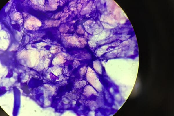
In some cases, your veterinarian may recommend a fine needle aspiration (FNA) test. For this, the vet will use a needle to perforate the mass and collect the material and/or cells within. FNA testing is useful for a wide variety of skin tumors, such as mast cell tumors, or subcutaneous (i.e. under the skin) tumors, such as lipomas in dogs. However, sometimes in the case of smaller or firm skin cysts (like sebaceous cysts), the aspirate may only yield normal skin cells, inflammatory cells, or cellular debris.
If FNA testing is inconclusive and the vet is concerned the mass could be something dangerous, he or she may recommend a biopsy. This is because biopsies, while more invasive, tend to provide more accurate results. Rather than pulling cells or debris from the mass like an FNA, a biopsy involves removing a small piece of tissue. The vet will then submit the tissue to a pathologist for further analysis.
Since some sebaceous cysts are very small, a cyst biopsy may mean surgically removing the cyst for further evaluation. As an added bonus, if the pathologist confirms the vet removed the entire cyst, the biopsy may be curative!
Pathologists may use the more correct term “keratin inclusion cyst,” “follicular cyst,” or “epidermal inclusion cyst” to describe what is commonly known as a sebaceous cyst. This is because true sebaceous cysts (i.e. those filled with sebum only) are relatively rare in veterinary medicine. Instead, the so called “sebaceous cysts” are also filled with keratin, which is the white or chunky part of the contents.
So don’t be surprised or concerned if you were expecting to see “sebaceous cyst” as the diagnosis and see one of these other terms instead. They are, for all practical purposes, the same thing.
Also, keep in mind that different types of lumps and bumps can look very similar. As a result, your dog’s FNA or biopsy may occasionally come back with an unexpected result. Benign tumors like hamartomas and sebaceous adenomas can look like sebaceous cysts but still carry a good prognosis. On the other hand, some cancerous bumps like squamous cell carcinoma may also mimic a sebaceous cyst. But a biopsy will reveal their true identity. No matter the diagnosis, your vet will be there to help you figure out the next steps—even if the plan looks different than you thought it would.

Why can sebaceous cysts become a problem?
Sometimes sebaceous cysts can be very “quiet” in nature. This means they are nothing more than a slight blemish on your dog’s skin. However, if the dog scratches the cyst or the groomer accidentally catches it with the clippers, bacteria and yeast on the skin can contaminate the site and cause infection. The area around an infected cyst may be red, inflamed, or have an unpleasant odor. If you notice any of these signs, your dog needs veterinary attention.
Also, as mentioned above, it is possible for sebaceous cysts to burst when the cyst becomes too full or does not have enough room to grow. Immediately after rupturing, you may notice a lot of discharge and/or bleeding from the site. It may also be painful or uncomfortable for your poor pup. You should make an appointment with your vet to address a ruptured or bleeding cyst as soon as you can. This is especially true if your dog is licking or biting the area or you notice signs your dog is in pain.
#2: Lipoma
Lipomas are one of the most common benign masses in dogs, and can affect any breed, although overweight or obese dogs, and older pets, are more likely to develop lipomas. These soft, rounded, fat-filled masses are typically found close under the skin, and are seldom painful. Some lipomas can develop under the muscles and feel firmer than subcutaneous lipomas, and may interfere with movement if they are poorly placed, such as around limbs. Most lipomas tend to be slow-growing, but, if they begin to grow quickly after a period of inactivity, surgical removal may be advised.
If your Dog has a Cyst, what your Veterinarian may do. Dr. Dan explains
You are stroking your pet’s silky fur while relaxing on the couch, when your hand stumbles across a small, firm lump. Instantly, your mind goes to “the big C”—cancer. While some lumps and bumps can indicate a cancerous tumor, not all masses are malignant. In fact, many lumps are benign and, although they may grow, are not likely to cause terminal illness. Before panicking about your furry pal’s lump, schedule an appointment with your Scripps Ranch Veterinary Hospital veterinarian for an accurate diagnosis. There’s an excellent chance your cherished companion’s bump is one of the following five most common benign masses.