What Are the Symptoms of Bone Cancer in Dogs?
The symptoms of your dogs bone cancer depend on where it develops. For osteosarcomas, the most frequent location is in one of your dogs limbs.Â
Osteosarcoma symptoms can include weakness, lameness, and pain in their limb. Osteosarcoma can also lead to a break in the bone at the spot where the tumor is located. Any broken bone in the limb of a large dog should be investigated as a possible case of osteosarcoma.Â
Axial osteosarcoma refers to a tumor located anywhere but the limbs, including the:Â
 In these cases, the tumor forms a large, solid mass that can also be quite painful.Â
The pain from tumors in both locations can lead to other symptoms, including:Â
Etiology and Risk Factors of Osteosarcoma
A major component of this disease in dogs, and possibly in people, appears to be genetic (i.e., heritable). Risk is most accurately defined by body mass, although there is a direct correlation with size as well. In children, osteosarcoma is frequently seen in kindreds with mutations of the retinoblastoma susceptibility gene (RB-1), and this risk is paternally imprinted. In dogs, there are clear breed predispositions. A recent study by Phillips and colleagues published in Genomics (Phillips et al., 2007) showed that the narrow heritability in Scottish Deerhounds was 0.69; in other words, almost 70% of the cause is due to heritable traits. Narrow heritability (h2) is the proportion of the total variability due to genetic factors. It is not surprising heritable factors account for a significant component of risk in Scottish Deerhounds; more than 15% of dogs from this breed die from osteosarcoma. The best-fit model for inheritance of the risk traits in Scottish Deerhounds was a Mendelian major gene with dominant expression. Furthermore, Comstock and colleagues (Comstock et al., 2006) reported at the 2006 Genes Dogs and Cancer meeting (Chicago, IL) there are 4 regions of the genome that appear to be associated with an increased risk of osteosarcoma in Rottweilers, another breed where risk appears greater than what would be attributable to size alone (incidence estimated at more than 12%).
Environmental factors that increase risk for osteosarcoma include rapid growth (therefore “large breed” puppy food has reduced levels of available energy to increase the time needed for these dogs to achieve their full size and mass potential), gender (the risk for males is 20 – 50% greater), and metallic implants to fix fractures. Chronic trauma and microscopic fractures have been proposed as risk factors, but this is difficult to prove conclusively. There was a study from David Waters group (Cooley et al., 2002), where survey data provided by owners showed an increase in risk to develop osteosarcoma in dogs that were spayed or castrated at an early age. The relative risk estimated from this study was as high as 4-fold higher for dogs neutered before one year of age than for intact dogs. Glickman’s group published similar data in 1998 based on analysis of cases in the Veterinary Medical Database (Ru et al., 1998). These studies generated significant debate and concern among veterinarians and owners. Nevertheless, the results have not been reproduced consistently in other large population studies (for example, Phillips et al and Scottish Deerhounds). While these results may have increased some owners’ reluctance to neuter or spay dogs, the possible 3-fold increase in risk of osteosarcoma in females should be placed in context of the 80 – 260-fold reduced risk of mammary cancer by early spaying, and the possible 4-fold increase in risk in males should be placed in context of behavioral problems, such as territorial aggression, roaming, marking behavior, and physiological problems such as prostatic hyperplasia and testicular cancers that appear more commonly (or exclusively) in intact male dogs.
There are three common histologic types of osteosarcoma: osteoblastic, where tumor cells produce large amounts of tumor osteoid; chondroblastic, where tumor cells produce cartilage (chondroid) in addition to some amount of tumor osteoid (without osteoid the diagnosis is chondrosarcoma); and fibroblastic, where tumor cells are predominantly fibroblasts and can produce both collagen and tumor osteoid. The disease is highly metastatic, with distant spread mostly to lungs and other sites in bone. Osteosarcoma can also metastasize to lymph nodes and intra-abdominal organs. The metastatic pattern is similar for dogs and humans.
Diagnosis is based on clinical signs, imaging, and biopsy. The clinical signs for appendicular osteosarcoma range from mild lameness with some evidence of pain to pathological fractures. The signs for axial and extraskeletal osteosarcoma are site-dependent. Imaging includes survey radiographs, and may be supplemented by magnetic resonance imaging (MRI) and/or computed tomography (CT) and nuclear scintigraphy. Imaging studies should include the primary tumor site and common sites of metastasis. Radiographic signs of osteosarcoma can range from severe lysis to severely sclerotic (increased density or hardening) lesions with new bone formation. There is usually loss of trabecular (internal) detail and indistinct demarcation of the tumor, associated soft tissue swelling, lysis of the outer boundary (cortex), and exuberant periosteal reactions that form the so-called “Codman’s triangle.” Although this is seen commonly, it is not always present and should not be considered the major determinant to make or rule out a diagnosis. Osteosarcoma rarely crosses joint space, except for an unusual type of necrotizing osteosarcoma of the tibia that is seen in Scottish Terriers and other smaller dogs.
Nuclear scintigraphy is very sensitive, but not specific to identify lesions associated with osteosarcoma, as any region of osteoblastic (bone growth or remodeling) activity will be identified (i.e., arthritis). Nuclear scintigraphy is useful to determine the extent of primary tumor involvement. Fine needle aspiration cytology is commonly used as an adjunct to confirm a radiographic diagnosis. Cytology alone is generally not sufficient to make a definitive diagnosis, but the presence of “flag cells” with eosinophilic material, granular cells, and variable cell size and shape can support the diagnosis. Definitive diagnosis requires a biopsy, which can be obtained through an open incisional biopsy, a trephine biopsy, or a Jamshidi bone marrow biopsy needle. The diagnostic accuracy is almost 100% for open biopsies, ~95% for trephines, and >90% for Jamshidi needle biopsies. Biopsies should be obtained from center of lesion, and if a limb-sparing procedure is elected, the surgeon performing the surgery should perform the biopsy whenever possible.
The pathologist will define the cell type (osteoblastic, chondroblastic, fibroblastic, mixed), grade (pleomorphism, proliferative fraction, etc.), and verify the presence of tumor osteoid, which is diagnostic. Other confirmatory tests can include immunohistochemistry, staining for osteocalcin, osteonectin, and alkaline phosphatase (ALP).
Treatment Options for Osteosarcoma in Dogs
Treatment recommendations for bone tumors depend on multiple factors andthe ultimate goal is to help improve your pet’s quality of life. Treatment canbe divided into two parts:
Bone Cancer in Dogs: Causes, Symptoms and And Answers to Frequently Asked Questions
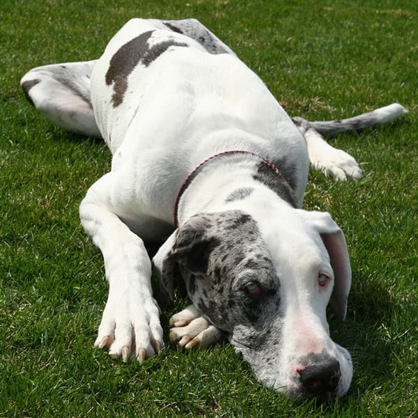
Osteosarcoma is a form of bone cancer in dogs that accounts for 85% of primary bone tumors diagnosed, making it the most common bone tumor. It is a highly aggressive tumor, characterized by local invasion and destruction of the bone as well as early metastasis (spread to other organs, the most common site of metastasis being the lungs). Osteosarcoma most commonly affects the limbs (or the appendicular skeleton) of large to giant breed dogs. It can also occur in other bones such as the skull, ribs, vertebrae, and pelvis (the axial skeleton) which are more common sites in smaller dogs.